
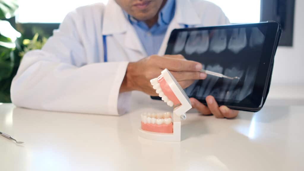
Dental X-ray frequency is a personalized diagnostic schedule based on your clinical risk of tooth decay and gum disease. While healthy adults typically require routine bitewing X-rays every 12 to 24 months, patients with active cavities or existing restorative work may require imaging every 6 to 12 months to ensure safety.
Digital Sensors vs. Daily Background Exposure
Medical literature supports that Wireless sensors consistently exhibit the ability to generate high-quality images, on par with or surpassing other digital devices [1] (Ardila et al., 2024). At Chatham Family & Implant Dentistry, we utilize digital sensors, which require up to 90% less radiation than traditional film. To put this in perspective, a routine set of four bitewing X-rays exposes you to roughly 0.005 millisieverts (mSv) of radiation. This is approximately the same amount of “background radiation” you naturally absorb from the environment during a short two-hour airplane flight or a single day spent in the North Carolina sun. We strictly adhere to the ALARA (As Low As Reasonably Achievable) principle, ensuring we capture only the images necessary to protect your health while keeping your exposure to a minimum.
Table that compares dental imaging modalities
Image type | Primary purpose | Common frequency | Clinical need |
Bitewings | Detects decay | 6-24 months | Routine monitoring |
Periapical | Views the entire tooth root | As needed | toothaches |
Full mouth series | Complete baseline of all teeth | 3-5 years | New patients |
3dCBCT scan | Bone density and nerve paths | 3-5 years | Implants and airway analysis |
Pediatric X-Ray Frequency: Why Children Need Faster "Snapshots."
Clinical evidence suggests that, depending on how a radiograph is taken, different tissues and anatomical structures may appear to have different attenuation and may even alter the size and location of organs [2] (Mowery et al., 2022). If you are a parent in Pittsboro, you may notice we recommend X-rays for your children more frequently than for yourself. This is because a child’s mouth is in a constant state of flux. Because primary (baby) teeth have thinner enamel than adult teeth, decay can travel from the surface to the nerve in as little as six months. Clinical guidelines suggest that children at high risk for cavities receive bitewing X-rays every 6 to 12 months until the decay is under control. For children at low risk, we may extend this to every 12 to 24 months. These routine images allow us to monitor jaw development, track the eruption of permanent teeth, and catch “interproximal” (between-teeth) cavities before they cause pain or premature tooth loss.
The "Caries Risk" Assessment: What Puts You in the High-Frequency Category?
Your X-ray schedule is based on your biology. During your exam, our dentists perform a Caries Risk Assessment to determine whether you need imaging every 6 months or every 2 years. You may be placed on a more frequent “High-Risk” schedule if you have:
- Receding Gums: Exposed tooth roots lack protective enamel and decay much faster than the crowns of your teeth.
- Existing Restorations: If you have multiple crowns, bridges, or large silver fillings, we need frequent X-rays to ensure bacteria are not underneath the dental work.
- Dry Mouth (Xerostomia): Saliva is your mouth’s natural cleaner.
- Deep Anatomy: Some patients naturally have deep grooves on their molars where plaque can hide, making them more susceptible to “hidden” decay.
Bitewings vs. Full Mouth Series: Understanding the 3-to-5-Year Rule
It is important to distinguish between the different types of diagnostic imaging we use. Bitewing X-rays are the routine images taken every year or two; they focus specifically on the crowns of your back teeth and the height of your supporting bone. However, a Full Mouth Series (FMX) or a 3D CBCT scan provides a comprehensive view of every tooth, the full root structure, and the surrounding jawbone. Because these images are so detailed, healthy adults typically need only an FMX or 3D scan every 3 to 5 years. This long-term baseline allows us to track silent changes in your jaw joint (TMJ) and screen for “asymptomatic” infections at the tips of your roots that classically would not show up on a standard bitewing.
The Financial ROI of Diagnostic Imaging: Why X-Rays Save You Thousands
From a “Preventive Planner” perspective, dental X-rays are the most cost-effective tool in modern medicine. A set of routine bitewings at our Pittsboro clinic is a small investment that identifies demineralization (early decay) while it is still reversible.
- The Proactive Path: If we spot a tiny shadow on an X-ray today, we can often treat it with a fluoride varnish or a small, affordable filling (approx. 150–300).
- The Reactive Path: If you skip the X-ray and the decay goes undetected for another year, it will likely reach the nerve. This turns a simple fix into a root canal and a porcelain crown, which can cost $1,500 to $2,500.
FAQS
1. Is it safe to get dental X-rays during pregnancy?
Yes. According to the ADA and the American College of Obstetricians and Gynecologists, dental X-rays are safe during pregnancy with appropriate shielding. However, unless you have an emergency or active pain, we often delay routine elective X-rays until after your baby arrives out of an abundance of caution.
2. Can I bring my X-rays from my previous dentist?
Absolutely. In fact, we encourage it! If you have had high-quality images taken within the last 6–12 months, we can often use those as your baseline. Our administrative team will even handle the legal record transfer for you to ensure we have the data we need before your first visit.
3. Why must the dentist just look at my teeth in conjunction with an X-ray?
Visual exams are great for the chewing surfaces, but they cannot see through the thick enamel to where teeth touch. Decay often starts in these “tight spots.” By the time a cavity is visible to the naked eye, it has usually progressed into the softer dentin, making the repair more complex and expensive.
About The Author
Dr. Colin Barbaro, DDS, provides comprehensive general and cosmetic dentistry at Azalea Dental in Wilmington, NC. With advanced surgical training from a VA hospital residency, he specializes in implant placement, TMJ therapy, restorative care, and endodontics. He is committed to delivering safe, evidence-based solutions that keep patients comfortable and informed at every step.
References
[1] Ardila, C. M., Vivares-Builes, A. M., & Pineda-Vélez, E. (2024). Image Quality, Radiation Dose, and Patient Comfort Associated with Wireless Sensors in Digital Radiography: A Systematic Review. Dentistry journal, 12(8), 267. https://doi.org/10.3390/dj12080267
[2] Mowery ML, Singh V. X-ray Production Technical Evaluation. [Updated 2022 Oct 17]. In: StatPearls [Internet]. Treasure Island (FL): StatPearls Publishing; 2026 Jan-. Available from: https://www.ncbi.nlm.nih.gov/books/NBK564332/